Curious to know how cell dances and glides in animal body? You need to hit here
Mumbai, April 27: We have often heard and learnt about the cells, which forms an integral part of the human body! But we have always been curious about what goes into a cell? Ain’t it true? Here is the video which will mesmerize you!
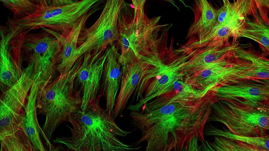
Yes, that’s a cell dancing and gliding in there! Although we've known for decades how cells move throughout organisms, this video is the first high-resolution, three-dimensional footage of that process as it's happening.
Those blue dots you can see the immune cell swallow up are particles of dextran, a sugary polysaccharide that's found in many substances - from wine and heart medication to dental plaque.
In addition to being absolutely mesmerizing, this footage is exciting because it's the first of its kind, done using a brand new type of microscopy.
Sure, we've seen cells under the microscope for hundreds of years. But when we try to get footage of them moving, the results are fuzzy. "This raises the nagging doubt that we are not seeing cells in their native state, happily ensconced in the organism in which they evolved," says team leader and physicist Eric Betzig, from the Howard Hughes Medical Institute in Virginia.
Even when we try to see just one cell at a time, the cells are blasted with intense light and our microscopes are too slow to follow all the action in 3D. "This also contributes to our fear that we are not seeing cells in their natural, unstressed form," says Betzig.
"It's often said that seeing is believing, but when it comes to cell biology, I think the more appropriate question is, 'When can we believe what we see?'"
To overcome this, Betzig and his team combined two microscopy technologies: adaptive optics, and lattice light sheet microscopy. Adaptive optics is the technology astronomers use to see distant celestial objects through Earth's rippling atmosphere.
In the case of imaging a living organism, this means shining a laser on whatever the researchers want to image, and using this to measure how much light distorts as it passes through surrounding cells and tissues.
The result is a totally clear image of a cell in its natural environment. The second technique - lattice light sheet microscopy - lets the researchers capture those super-clear images in real time.
It involves rapid and repeated sweeps of ultra-thin sheets of light, which creates a series of 2D images that can be built into a moving high-res 3D picture, all without frying the cell or interfering with its activity.
The result is something scientists have wanted for years - bright, clear, and vibrant images of our cells in action under the microscope.